The Institute of Applied Ophthalmology (IOBA) of Valladolid (North western Spain) and the Institute of Microelectronics of Barcelona (IMB-CNM-CSIC) (North eastern Spain) have developed a sensor capable of diagnosing, in live, some alterations of the cornea from the impedance measurement, ie the resistance of the tissues to the passage of electricity.
This is the main contribution of the doctoral thesis of the researcher of the IOBA, Ana del Rio, which is in its final phase and she investigates how they affect the contact lenses to the cornea, using this device, as reported Dicyt.
The objective of this work is to put into clinical practice new non-invasive diagnostic methods of the cornea. This organ is one of the most important parts of the eye, consisting of five layers: epithelium, Bowman's membrane, stroma, Descemet's membrane and endothelium.
Besides protecting other ocular structures, it helps the eye to focus images on the retina. Currently, there is a lack in terms of non-invasive diagnostic devices of the cornea, which limits its study to certain conditions or how they affect the use of contact lenses in some situations. "In the cornea, the layer bounded on the outside (the epithelium) and bounded on the inside (the endothelium) have some overlap but they have to be enough strong to form a barrier to protect the cornea from abroad, for the epithelium, and to protect it from excessive water does not flow from the anterior chamber, so that occurs a corneal edema, in the case of endothelium”, the researcher explained.
Thus, it is known how a contact lens affects the permeability of these two layers, the epithelium and endothelium. As indicated by del Rio, there is a more or less famous technique in clinical practice, lafluorofotometría, which allows to study the cornea but, notes, "it has several downsides", today.
Between them that the measure in the patient lasts several hours and having low reliability, because "there are several studies and it seems that no one agrees on what actually happens with the layers of the epithelium and endothelium, when a lens is placed contact" , rec In the absence of tools to objectively measure and direct the permeability of these layers, we used the equipment developed by the Institute of Microelectronics of Barcelona, a noninvasive sensor that rests on the corneal surface for a few seconds and measures the impedance, recalls the Doctor.
In the absence of tools to objectively measure and direct the permeability of these layers, we used the equipment developed by the Institute of Microelectronics of Barcelona, a noninvasive sensor that rests on the corneal surface for a few seconds and measures the impedance.
First, it has made a preliminary experimental study in rabbits with contact lenses and the results have been positive, "which has led us to use the device to measure permeability in the human cornea".
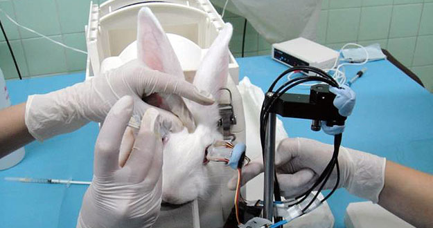
The researchers tested the ability of the device diagnosed in laboratory animals
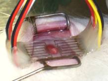
Researchers testing the nanolectrodos in a rabbit cornea
The main contribution, that the thesis tryes doing is to make sure that the device is safe for use in humans and, secondly, to detect whether the measures actually hits the sensor indicate how the permeability of subjects carrying contact lenses . To do this, they have conducted a pilot study in which 10 subjects were included.
"We have randomly placed contact lenses of different permeability, high and low, because, according to her, the changes, that will occur on the epithelium and endothelium, will be different", said and stressed that they were intended to check whether capable of detecting the changes in the function of the contact lens, that has been assigned to each patient. These ten subjects took contact lenses for a week and the permeability measurements, with this equipment, were performed before putting the contact lenses (baseline measures compared with other results), after three days of taking the lenses and after one week.
The sensor has no damage to the cornea of patients. Regarding the first objective of the study: to assess whether the method is safe, del Rio said: "We can say that it is, because there were no primary or secondary damage to the cornea of patients to apply the sensor".
As for the effectiveness of the device, she said: "We can not say it categorically, as the number of subjects included in the study was quite small and it should have also improve the repeatability of the team, so it will take more research".
Looks interesting this advance. Given that the eye is a complex organ, any progress, it seems to me a miracle.
Till soon, kind regards,
Luis.
Sponsored by Costaluz Lawyers.
Please click down here:
